Issued Date: 2019/5/14
Issued By: iST
Can I use 3D X-ray to check internal structure and defects in my non-IC or non-metal products?
Being a non-destructive inspection mechanism, 3D X-ray inspection is something frequently presented in iST Tech Classrooms. This tool first presents samples in 3D images, then uses CT slice images to identify defects in the internal structure, material, or packaging of the sample.
iST Tech Classroom used to focus on using 3D X-ray to test IC or metal samples (Read on: What 3D X-ray upgrade can do for you?). In fact, 3D X-ray are also ideal for non-destructive inspection over 3D IC (Read on: 3 Steps to identify the causes of 3D-IC failures), MEMS, and even PCB, PCBA, lithium battery/engineering Plastics, and systems as required by you. (Read on: When is the Best Time to Identify Defects with 3D X-ray?)
In iST Tech Classroom this month, we’ll shift our focus to using 3D X-ray for checking non-IC and PCBA samples.
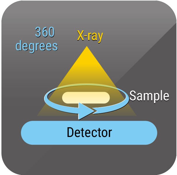
High Resolution 3D X-Ray Microscope is a non-destructive method employed with an optical microscope to enhance the magnification, which uses 2D X-ray tomography to take sample images at all angles while turning the fixed sample 360 degrees. Physical images of the tested object are then constructed by computer operation.
Case 1: Identify Internal Defects in Lithium Battery with 3D X-ray
3D X-ray can view defects within a battery without damaging it. Most lithium batteries are made with positive electrodes, negative electrodes, electrolytes, and a separator film. These are assembled by stacking or wrapping the positive and negative electrodes in an electrolyte filled shell with the separator film serving to isolate both electrodes and ensure free ion flow in between. In spite of its irrelevance with electrochemical reaction structure and properties of the separator film, the performance of the lithium battery would be hampered. In these cases, we found cracks and particles in the electrode plating of the lithium battery (see Fig. 1 and Fig. 2).
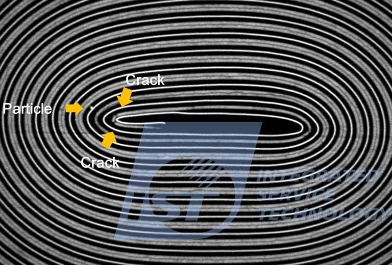
Figure 1: Positive/negative electrodes in battery bent to crack and particles found in between electrodes and separation film.
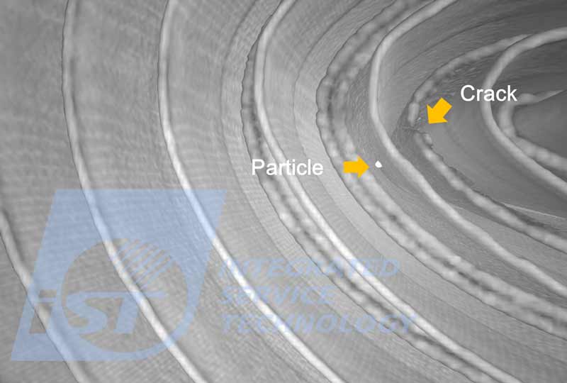
Figure 2: Internal 3D image of battery suggests the existence of cracks and particles.
Case 2: View Internal Structure of Engineering Plastics with 3D X-ray
The engineering plastics PC, PMMA, ABS, POM, PBT, and TPU among other engineering plastics are used as material for mechanic engineering structure. This precision injection molded medical product, requiring no deformation under high temperature, is made of PSUs featuring heat-resistance and dimensional stability. With the help of 3D X-ray, we found there are cracks and voids in its structure (see figure 3 and 4).

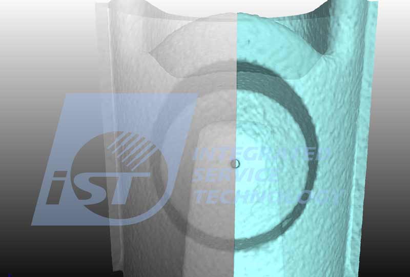
Figure 3 (left) and Figure 4 (right): medical grade plastic goods mandate dimensional accuracy. The 3D X-ray tool is ideal for viewing its internal structure and mechanical precision. Figure 3 suggests any defects in the nozzle and diameter measurements while figure 4 shows its structure with a 3D model.
Case 3: View Internal Structure of Optical Lens Mounting in Consumer Electronics
Optical lenses mounted on most consumer electronics may come with elements made by glass or PC/PMMA. Each optical lens is designed to consist of about 5-6 lenses. The quality of each of them may be hampered by initial design, mold accuracy and final assembly. The latter, in turn, is subjected to lenses alignment as well as precision of optical axis focus compliance with a given MTF (modulation transform function). The yield of module assemblers may vary greatly with skill and complexity. As a result, manufacturers are required to look into internal structure during assembling process. In this case, we use 3D X-ray to inspect internal structure of an assembled lens. Figure 5 shows whether there are defects in the assembly process between each lens and the lens group.
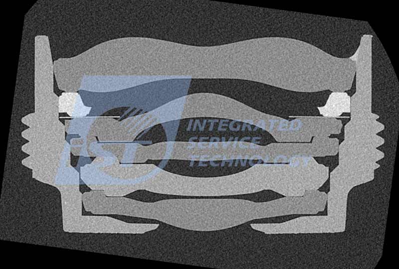
Figure 5: An optical lens is composed of 5 lenses. The assembling process should ensure each lens is aligned straight and the optical axis is accurate. 3D images enable clear views of the internal structure.
Case 4: Identify Internal Defects in PCBA with 3D X-ray
Amid the continuously growing PCB (Printed Circuit Board) industry, the appliance industry keeps on imposing heavier demands on PCBs in terms of material, layers, accuracy, and BLR (Board Level Reliability). 3D X-ray can be employed to identify defects in PCBA (Printed Circuit Board Assembly) including blind/buried via open (BVH open), crack, particle and burnout. Figure 6-8 suggests BVH opening and traces of burnout found in PCBA with 3D X-ray.
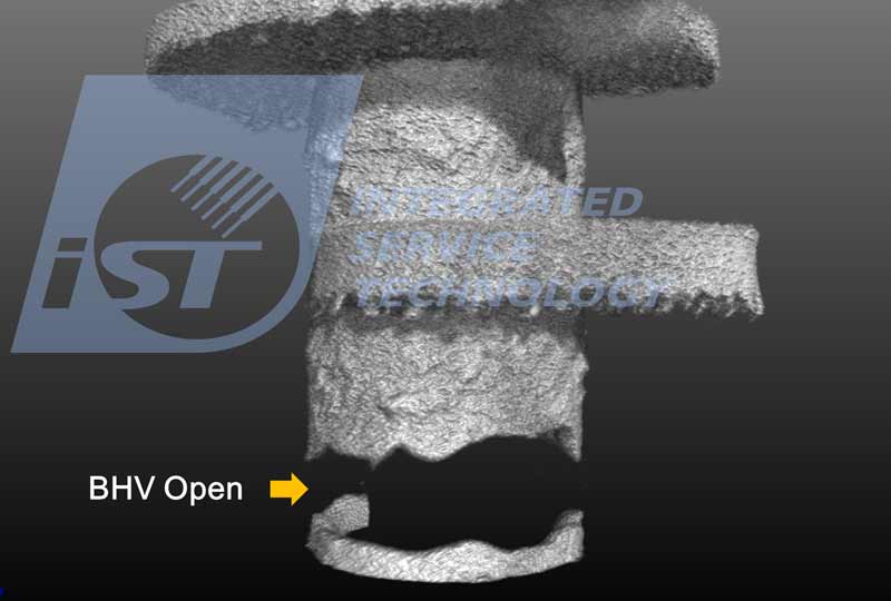
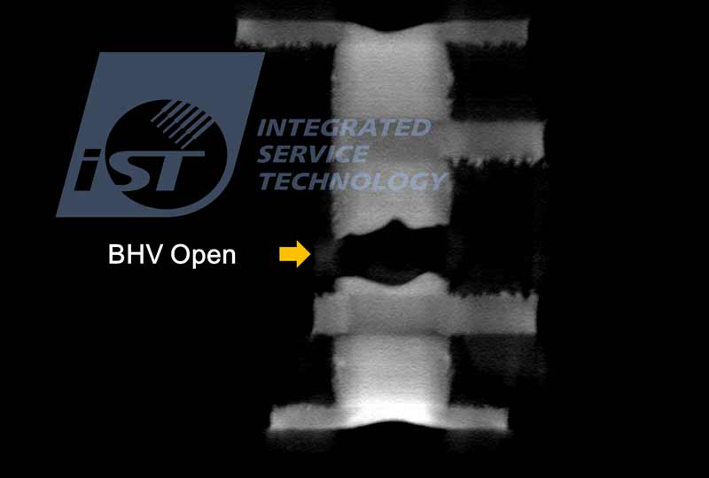
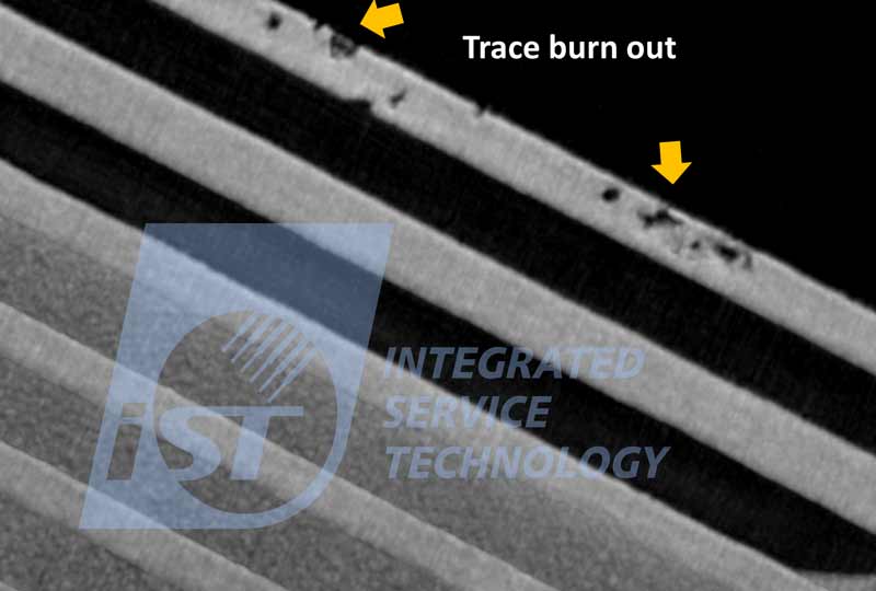
Figure 6(left), Figure 7(center), Figure 8(right): Defects found in PCBA and substrate processes include voids and blind voids defects (figure 6-7); poor circuit process and burnout (figure 8); defects including bridging, open circuit, board burnout, and analysis over wiring of individual layers in multi-layer boards can be viewed with 3D X-ray.
Case 5: The Maximum Sample Size for 3D X-ray Inspection
Through the recent cooperation with YXLON, a German company, iST now can scan samples of size up to 30*50cm at high power up to 225kV/280W by YXLON FF35 3D CT X-ray equipment.
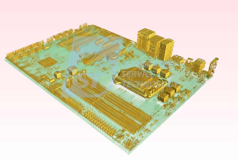
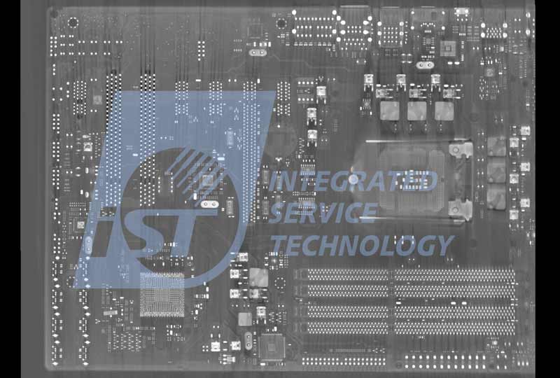
Figure 9-10: 3D X-ray image of computer main board at size of up to 22*30cm, the FF35 3D CT X-ray equipment can take pictures of big samples in one shot while the quality of the image remains acceptable in spite of big differences in density.
Should you have any requirements for sample defects, please call Rita Chen at +886-3-579-9909 Ext. 6571 or Kevin Chang at Ext. 6580 or send an email to web_nde@istgroup.com.